
Carotid Stenting / Intracranial Stenting
Home > Carotid Stenting / Intracranial Stenting
Carotid Stenting / Intracranial Stenting
Overview
Carotid and intracranial stenting are advanced, minimally invasive procedures used to treat narrowed or blocked arteries in the neck (carotid arteries) or inside the brain (intracranial arteries). These procedures help restore normal blood flow, prevent stroke, and protect vital brain functions.
Under the expert care of Dr. Gaurav Goel, a leading Neurointervention and Endovascular Neurosurgeon, patients receive cutting-edge treatment with precision, safety, and personalized care.
What Is Carotid Stenting?
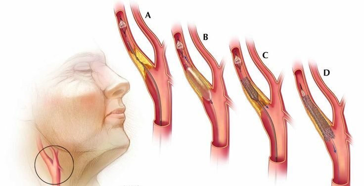
The carotid arteries supply blood to the brain. When these arteries become narrowed due to atherosclerosis (plaque buildup), it increases the risk of stroke.
Carotid stenting involves placing a small, expandable metal mesh tube (stent) inside the artery to open the blockage and keep the artery open, allowing blood to flow freely to the brain.
What Is Intracranial Stenting?
Intracranial stenting is performed inside the brain to treat narrowed or blocked intracranial arteries that may lead to recurrent strokes. This advanced technique helps improve blood circulation in brain regions at risk and is typically considered when medical management alone isn’t sufficient.
Conditions Treated
Carotid artery stenosis
Intracranial arterial stenosis
Transient Ischemic Attack (TIA)
Ischemic stroke caused by arterial narrowing
Atherosclerosis affecting brain or neck arteries
Symptoms That May Indicate the Need for Evaluation
Sudden weakness or numbness on one side of the body
Difficulty speaking or understanding speech
Vision loss or blurring in one or both eyes
Dizziness, loss of balance, or coordination problems
Episodes of temporary paralysis (mini-strokes)
If you experience any of these warning signs, immediate medical consultation is crucial to prevent a major stroke.
Procedure Overview
Diagnosis & Imaging – The patient undergoes advanced brain and neck imaging such as CT Angiography, MR Angiography, or Digital Subtraction Angiography (DSA) to locate the blockage.
Local Anesthesia – The procedure is performed under local anesthesia with minimal discomfort.
Catheter Insertion – A thin catheter is inserted through the groin or wrist and guided to the affected artery using imaging guidance.
Balloon Angioplasty & Stent Placement – The narrowed section is widened using a balloon, and a stent is carefully deployed to keep the artery open.
Post-Procedure Care – Patients are monitored for a short period and can usually resume normal activities within a few days.