AVM Embolization Treatment
Home > AVM Embolization
AVM Embolization Treatment in Delhi NCR — By Dr. Gaurav Goel
Overview
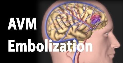
Arteriovenous Malformation (AVM) Embolization is a minimally invasive procedure performed to block abnormal blood vessels in the brain or spinal cord that connect arteries and veins directly, bypassing capillaries. These abnormal tangles of blood vessels can rupture and cause bleeding in the brain (hemorrhage), seizures, or neurological problems if left untreated.
Dr. Gaurav Goel, one of the leading Neurointerventional Surgeons in Delhi NCR, specializes in performing AVM Embolization using the latest image-guided techniques to ensure precision, safety, and faster recovery.
What is an AVM?
An Arteriovenous Malformation (AVM) is a congenital defect in which arteries and veins are abnormally connected. This causes:
High blood flow directly from arteries to veins
Weakening of vessel walls
Risk of rupture and bleeding
AVMs can occur in the brain, spine, or other parts of the body, but cerebral (brain) AVMs are the most serious due to the risk of stroke and brain damage.
What is AVM Embolization?
AVM Embolization is an advanced endovascular treatment performed through a small incision in the groin or wrist. A thin catheter is guided through blood vessels under fluoroscopic (X-ray) guidance to reach the AVM site. Once positioned, a special embolic material (such as glue, coils, or Onyx) is injected to block the abnormal vessels and reduce blood flow to the AVM.
Goals of AVM Embolization
Prevent AVM rupture and brain hemorrhage
Reduce AVM size before surgery or radiosurgery
Alleviate symptoms like headaches, seizures, or neurological deficits
Provide standalone treatment in select cases
Who Needs AVM Embolization?
Dr. Gaurav Goel recommends AVM Embolization for patients with:
Diagnosed brain or spinal AVM
Symptoms such as seizures, headaches, or weakness
History of intracranial bleeding due to AVM rupture
Large or complex AVMs that require staged treatment
Procedure Steps
Pre-Procedure Evaluation:
MRI, CT angiography, or digital subtraction angiography (DSA) is performed to map the AVM.Catheter Navigation:
A microcatheter is guided through arteries to the AVM.Embolization:
Embolic agents (glue, Onyx, or coils) are injected to block abnormal vessels.Post-Procedure Monitoring:
Patients are observed in a neuro-ICU for 24–48 hours for recovery and follow-up imaging.