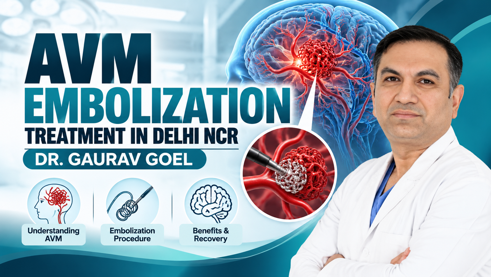
An arteriovenous malformation (AVM) is an abnormal connection between arteries and veins that bypasses the normal network of tiny blood vessels called capillaries. When an AVM develops in the brain, it can disrupt normal blood flow and, in some cases, cause bleeding, seizures or other neurological complications.
AVM embolization is a minimally invasive, image-guided procedure that involves introducing a specialised material into the abnormal blood vessels to reduce or block blood flow through the AVM. Depending on the AVM’s size, location and structure, embolization may be used before surgery or radiosurgery, or as a standalone treatment in carefully selected cases.
For patients seeking AVM embolization treatment in Delhi NCR, an accurate diagnosis and an individualised treatment plan are essential. Dr. Gaurav Goel is a neurointervention specialist at Medanta, Gurugram, with expertise in treating vascular conditions of the brain and spine, including AVM embolization.
What Is a Brain Arteriovenous Malformation (AVM)?
A brain AVM is a tangle of abnormal blood vessels in which arteries connect directly to veins without the normal capillary network in between.
Normally, arteries carry blood under relatively high pressure towards the body’s tissues. Capillaries allow oxygen and nutrients to pass into those tissues before blood returns through the veins. In an AVM, this normal process is bypassed, creating an abnormal high-flow connection.
Some AVMs remain undetected until an imaging test is performed for another reason. Others cause symptoms when they bleed, affect nearby brain tissue or interfere with normal blood flow.
Potential complications include:
- Bleeding in or around the brain
- Seizures
- Headaches
- Weakness or numbness
- Problems with speech, vision or coordination
- Other neurological deficits
Not every AVM will rupture, and the risk varies considerably between individuals. A specialist assessment helps determine whether treatment is appropriate and which approach may offer the best balance of benefits and risks.
What Is AVM Embolization Treatment?
AVM embolization, also known as endovascular embolization, is a procedure performed from inside the blood vessels.
Rather than requiring an open operation to reach the abnormal vessels, the treating specialist guides a thin catheter through the arterial system towards the AVM under X-ray guidance. A smaller microcatheter is then positioned at the intended treatment site, and an embolic material is delivered to block selected abnormal channels.
Materials may include specialised liquid embolic agents or other devices, depending on the AVM’s anatomy and the treatment plan.
The main objectives of AVM embolization may include:
- Reducing blood flow through the abnormal vessels
- Treating specific high-risk parts of an AVM
- Preparing the AVM for surgical removal
- Supporting subsequent stereotactic radiosurgery
- Completely closing the AVM in selected cases where this can be achieved safely
Important: Embolization does not automatically eliminate every AVM. The treatment team must confirm the degree of closure and determine whether additional treatment or follow-up imaging is needed.
Who May Need AVM Embolization?
AVM embolization may be considered for patients with a diagnosed brain or spinal AVM when the potential benefits of intervention outweigh its risks.
A neurointervention specialist may evaluate embolization in situations such as:
1. AVM Associated With Bleeding
An AVM that has ruptured requires urgent medical evaluation. Depending on the bleeding, anatomy and clinical condition, embolization may be used to address the source of bleeding or prepare the AVM for definitive treatment.
2. Complex or Large AVMs
Some AVMs are difficult to treat using a single procedure. Embolization may be performed in stages or used to reduce blood flow before surgery or radiosurgery.
3. AVMs With Seizures or Neurological Symptoms
Patients experiencing seizures, neurological deficits or other symptoms may need a detailed assessment to establish whether the AVM is responsible and whether intervention is appropriate.
4. AVMs Requiring Preoperative Treatment
In selected patients, embolization can reduce blood flow through parts of an AVM before microsurgical removal. This may help the surgical team plan the operation, although the benefit depends on the individual vascular anatomy.
5. Selected AVMs Suitable for Embolization Alone
Some AVMs can potentially be treated completely through an endovascular approach. This depends on their structure, location, feeding arteries and draining veins, as well as the likelihood of safely achieving complete closure.
The final treatment decision should be made by an experienced multidisciplinary team.
How Is AVM Embolization Performed?
The procedure is planned using detailed imaging and performed in a specialised angiography suite.
Step 1: Detailed Diagnostic Evaluation
The specialist reviews the patient’s symptoms, medical history and previous scans. Imaging may include MRI, CT angiography and digital subtraction angiography (DSA).
DSA provides detailed information about the AVM’s feeding arteries, abnormal vascular connections and draining veins. It is particularly useful for treatment planning.
Step 2: Preparation for the Procedure
The medical team explains the procedure, its potential benefits and risks, and the planned approach. Blood tests, medication review and anaesthesia assessment may be required.
The procedure is commonly performed under general anaesthesia, although the approach depends on the case and the treating team’s protocol.
Step 3: Catheter Navigation
A small arterial access point is created, commonly in the groin or wrist. A catheter is guided through the blood vessels under X-ray imaging towards the arteries supplying the AVM.
Step 4: Delivery of the Embolic Material
A microcatheter is positioned close to the intended treatment area. The specialist delivers the selected embolic material to block targeted abnormal vessels.
The procedure requires precision because normal brain tissue depends on an adequate blood supply. The objective is to treat the intended abnormal vascular connections while minimising the risk of damage to healthy vessels.
Step 5: Angiographic Assessment
Further angiographic images are taken to evaluate the effect of embolization and determine whether additional treatment is necessary.
Depending on the results, the team may plan another embolization session, surgery, radiosurgery or follow-up imaging.
Step 6: Post-Procedure Monitoring
After embolization, the patient is monitored for neurological changes, bleeding at the access site and other complications. The length of hospital stay depends on the complexity of the procedure and the patient’s condition.
What Are the Benefits of AVM Embolization?
When appropriately selected, AVM embolization can offer several potential benefits.
Minimally invasive access: The procedure is performed through blood vessels, avoiding the need for a traditional open craniotomy solely to access the AVM.
Targeted treatment: Imaging allows the specialist to guide the catheter towards specific abnormal vascular connections.
Support for combined treatment: Embolization may help prepare selected AVMs for microsurgery or radiosurgery.
Potential for complete closure in selected cases: Some AVMs may be completely treated through embolization, although this cannot be guaranteed.
Individualised treatment planning: The procedure can be tailored to the AVM’s anatomy and the patient’s clinical needs.
The actual benefits depend on the AVM’s characteristics, the treatment objective and the risks of intervention.
What Are the Risks of AVM Embolization?
Although AVM embolization is minimally invasive, it is a complex neurovascular procedure and carries important risks.
Potential complications include:
- Stroke or reduced blood supply to healthy brain tissue
- Bleeding or rupture of abnormal vessels
- Damage to normal blood vessels
- Seizures
- New or worsening neurological deficits
- Swelling or haemorrhage following treatment
- Complications related to the catheter access site
- Reactions to contrast material
- Complications associated with anaesthesia
- Incomplete closure or the need for further treatment
The level of risk depends on the AVM’s location, size, vascular structure and the planned intervention. A detailed discussion with the treating specialist is essential before proceeding.
AVM Embolization vs Surgery vs Radiosurgery
There are several treatment approaches for brain AVMs. Each serves a different purpose, and some patients benefit from a combination of treatments.
| Treatment | How it works | When it may be considered |
|---|---|---|
| AVM embolization | Uses a catheter to deliver material that blocks abnormal vessels | Selected AVMs, preoperative treatment or part of a combined plan |
| Microsurgical resection | Surgically removes the AVM through an opening in the skull | Selected AVMs that can be removed with an acceptable level of risk |
| Stereotactic radiosurgery | Uses focused radiation to damage abnormal vessels so they gradually close | Selected AVMs, particularly those suited to focused radiation |
| Observation | Monitors the AVM through clinical assessment and imaging | Selected cases where intervention may pose greater risk than observation |
Radiosurgery does not close an AVM immediately; obliteration can take months or years, during which bleeding risk may remain. Similarly, embolization may not provide complete closure in every case.
The best approach depends on the AVM’s anatomy, previous bleeding, symptoms, patient age, overall health and the risks of treatment.
Recovery After AVM Embolization
Recovery varies according to the complexity of embolization, the patient’s neurological condition and whether additional treatment is needed.
Immediately After the Procedure
The patient is monitored in a recovery area or intensive care setting as clinically indicated. The team checks neurological function, vital signs and the arterial access site.
Some patients may experience temporary discomfort, fatigue or a headache. New or severe symptoms require prompt assessment.
During the First Few Days
Hospital observation may be needed, especially after complex procedures or when the AVM has previously bled.
The medical team provides instructions about medications, activity, wound care and follow-up. The timing of discharge depends on the patient’s condition rather than a fixed schedule.
Follow-Up and Long-Term Monitoring
Follow-up imaging is important to assess how much of the AVM remains and whether further treatment is required.
Even when symptoms improve, patients should not assume the AVM has been completely eliminated. The specialist will determine whether angiographic confirmation or continued surveillance is necessary.
Why Choose Dr. Gaurav Goel for AVM Embolization in Delhi NCR?
AVM treatment requires detailed knowledge of neurovascular anatomy, advanced imaging and careful treatment planning.
Dr. Gaurav Goel is the Senior Director and Head of Neurointervention Surgery at Medanta, Gurugram. His training includes a DM in Neuroradiology from NIMHANS, Bengaluru, and fellowship training in interventional neuroradiology at the Montreal Neurological Institute, McGill University, Canada.
His areas of clinical expertise include AVM embolization, intracranial aneurysm treatment, stroke interventions and other neurovascular procedures.
Patients considering AVM embolization in Delhi NCR should seek a consultation to review their imaging, understand the available treatment options and discuss the potential benefits and risks of each approach.
An individualised treatment plan should clarify:
- Whether embolization is appropriate for the specific AVM
- Whether the procedure is intended as a standalone treatment or part of a combined plan
- The expected degree of AVM closure
- Potential complications and alternative treatments
- The follow-up imaging schedule
When Should You Seek Emergency Medical Care?
A brain AVM can sometimes cause bleeding that requires urgent treatment.
Seek emergency medical attention if you or someone nearby develops:
- A sudden, severe headache
- A seizure, particularly a first seizure
- Sudden weakness or numbness
- Difficulty speaking or understanding speech
- Sudden vision or balance problems
- Loss of consciousness or confusion
These symptoms can indicate a serious neurological emergency. Do not wait for a routine appointment or attempt to arrange an outpatient consultation first.
Frequently Asked Questions About AVM Embolization
1. What is AVM embolization treatment?
AVM embolization is a minimally invasive procedure in which a specialist guides a catheter through the blood vessels and delivers an embolic material to block targeted abnormal connections between arteries and veins.
2. Is AVM embolization a brain surgery?
It is an endovascular procedure rather than traditional open brain surgery. It involves accessing the blood vessels through a small arterial puncture and guiding catheters to the AVM under imaging guidance.
3. Can AVM embolization completely cure a brain AVM?
It can completely close some AVMs in selected cases, but many require additional treatment. Follow-up imaging is needed to establish whether complete obliteration has been achieved.
4. Is AVM embolization safe?
The procedure can be appropriate for selected patients, but it carries risks, including stroke, bleeding and neurological injury. The risks and expected benefits must be assessed individually by the treating specialist.
5. How long does AVM embolization take?
The duration varies according to the AVM’s size, location, complexity and the number of vessels requiring treatment. Some AVMs need multiple sessions rather than a single procedure.
6. Is embolization always required before AVM surgery?
No. Embolization is useful in selected cases, but it is not necessary for every AVM. Some can be treated with surgery or radiosurgery, while others may be monitored. The decision depends on the vascular anatomy and the treatment plan.
7. What is the difference between AVM embolization and radiosurgery?
Embolization blocks targeted abnormal vessels through a catheter-based procedure. Radiosurgery uses focused radiation to make the abnormal vessels close gradually over time. The procedures have different indications and may sometimes be used together.
8. Can a brain AVM return after embolization?
An AVM may remain partially open or recur after treatment in some cases. Follow-up imaging helps determine whether complete closure has occurred and whether further treatment is required.
9. Which specialist treats brain AVMs in Delhi NCR?
Brain AVMs are evaluated by specialists such as neurointerventionalists and vascular neurosurgeons. A multidisciplinary team may include neuroradiologists, neurologists and neurosurgeons to determine the most appropriate approach.
10. Where can I consult Dr. Gaurav Goel for AVM embolization?
Dr. Gaurav Goel practises at Medanta, Gurugram. Visit his official website to review his profile and contact information and to enquire about an appointment for AVM evaluation.
Conclusion
AVM embolization is an important treatment option for selected patients with brain or spinal arteriovenous malformations. By targeting abnormal blood vessels through a catheter-based approach, it may reduce blood flow through an AVM, support other treatments or completely close selected lesions.
However, treatment decisions must be based on detailed imaging and an assessment of the potential risks and benefits. Some AVMs require combined treatment, while others may be better managed with a different approach or careful monitoring.
If you are looking for AVM embolization treatment in Delhi NCR, consult Dr. Gaurav Goel to discuss your diagnosis, available treatment options and the next steps appropriate for your condition.
Website: https://drgauravgoel.org/